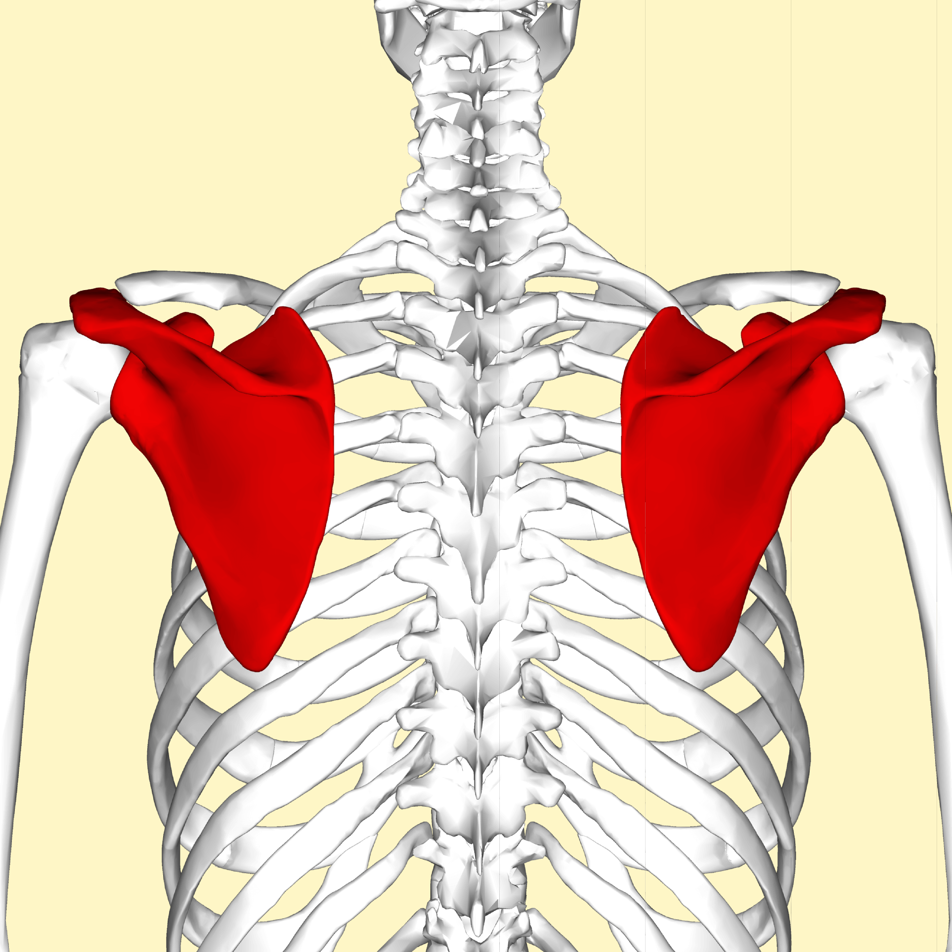

The subscapular fossa is separated from the vertebral border by smooth triangular areas at the medial and inferior angles, and in the interval between these by a narrow ridge which is often deficient. The lateral third of the fossa is smooth and covered by the fibers of this muscle. The ridges give attachment to the tendinous insertions, and the surfaces between them to the fleshy fibers, of the Subscapularis. The medial two-thirds of this fossa are marked by several oblique ridges, which run lateralward and upward. Pectoral girdle - front Human arm bones diagram Diagram of the human shoulder joint Left scapula. The costal or ventral surface presents a broad concavity, the subscapular fossa. The following muscles attach to the scapula: The scapula also articulates with the clavicle, via the acromion process (the acromioclavicular joint). This forms the socket that the head of the humerus articulates with. Near the base of the coracoid process, so also on the lateral angle, there is a depression called the glenoid cavity. The end of this hook is the site of attachment of many muscles, such as the coracobrachialis muscle. For humans and carnivores and bovinae the spina runs into a forward pointing hook called acromion, which continues past the main part of the bone.Īnother hook-like projection comes off the lateral angle of the scapula, and is called the coracoid process. After this peak the spina scapulae steeply decays in height. It begins flat at the base of the shoulder bone, ascends in distal direction to its peak at about the middle of the scapula, this peak is called tuber scapulae. This projection is called the spine of the scapula. The posterior surface of the scapula is divided by a bony projection, the spina scapulae (opposite to the fossa subscapularis) into the supraspinous fossa and the infraspinous fossa. The anterior (front) side of the scapula shows the fossa subscapularis (subscapular fossa) to which the subscapularis muscle attaches. It has two surfaces, three borders, and three angles.


 0 kommentar(er)
